|
|
Probe Detection |
|
|
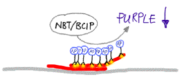
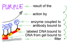
Immune detection of the probeDigoxigenin (DIG), which is used to label the probe, is widely used for immune detection. This plant steroid molecule is highly antigenic. Because it is found exclusively among foxglove (Digitalis) plants, antibodies directed against it will not cross-react with antigens from other organisms. So, anti-digoxigenin can be used to very specifically detect the digoxigenin on a hybridised blot. To be able to visualise the (polyclonal) DIG antibodies when they are bound to the digoxigenin, they have been covalently linked to the enzyme alkaline phosphatase (AP). When the antibody/enzyme conjugate is used to bind the digoxigenin label on the blot, an immune complex is produced. This step is again preceeded by a blocking procedure, this time to block any nonspecific antibody binding sites on the blot. In a subsequent step, the antibody/digoxigenin complex is detected by two color-forming reactions. First, alkaline phosphatase catalyses the removal of a phosphate group from 5-bromo-4-chloro-3-indoylphosphate (BCIP); the resulting oxidation product dimerizes to form an indigo precipitate. Hydride ions released during dimerization reduce a second compound, nitro blue tetrazolium (NBT), to form a purple precipitate. The amount of the two precitates deposited on the membrane is proportional to the enzymatic activity and hence to the mass of target DNA present. So, a more intense blue/purple color indicates more target DNA on the blot. See the pictures below
The purple precipitate is the result of ...
|